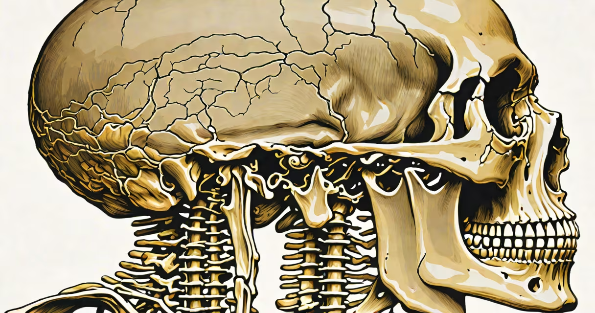
The axial skeleton meaning refers to the central core of the human body's skeletal structure, comprising 80 bones that form the upright axis from the skull to the pelvis. It includes the skull, vertebral column, sternum, and ribs, and serves as the primary support for the head, neck, and trunk while protecting important organs such as the brain and heart.
This structure is distinct from the appendicular skeleton, which includes the limbs and girdles. The axial skeleton accounts for about half the total bone count in the adult human body and forms the foundation for posture, balance, and movement coordination. Its design prioritizes protection and stability over mobility, unlike limb bones that emphasize motion.
According to Gray's Anatomy (41st edition), the axial skeleton "provides structural integrity and houses delicate neural and circulatory systems." This role is critical during daily activities-from breathing and speaking to maintaining upright stance under gravitational load.
The axial skeleton functions through a combination of rigid bony structures and flexible joints. The skull encases the brain within the cranial cavity, a bony vault that resists impact and maintains intracranial pressure. The vertebral column, composed of 33 vertebrae divided into cervical, thoracic, lumbar, sacral, and coccygeal regions, supports the upper body and allows controlled bending and twisting.
Each vertebra contains a vertebral foramen that aligns to form the spinal canal, housing the spinal cord. Intervertebral discs between adjacent vertebrae act as shock absorbers, distributing mechanical stress during walking, lifting, or jumping. These discs are made of fibrocartilage and consist of a gel-like nucleus pulposus surrounded by a tough annulus fibrosus.
The rib cage, formed by 12 pairs of ribs connected to the sternum via costal cartilages, protects the heart and lungs. During inhalation, the diaphragm contracts and the rib cage expands upward and outward-this movement increases thoracic volume and draws air into the lungs. Without the structural rigidity of the axial skeleton, this process would be mechanically inefficient.
The axial skeleton consists of five major groups: skull bones, auditory ossicles, hyoid bone, vertebral column, and thoracic cage. Below is a breakdown of key elements:
| Bone Group | Number of Bones | Primary Function |
|---|---|---|
| Skull (cranial & facial) | 22 | Protects brain; provides attachment points for muscles involved in chewing and facial expression |
| Vertebral column | 26 (including sacrum and coccyx) | Supports head and trunk; protects spinal cord; enables limited flexion and extension |
| Rib cage | 24 (12 pairs) | Shields heart and lungs; assists in ventilation via expansion and contraction |
| Hyoid bone | 1 | Supports tongue and larynx; anchors muscles involved in swallowing and speech |
| Auditory ossicles | 6 (3 in each ear) | Transmit sound vibrations from eardrum to inner ear |
The hyoid bone is unique-it does not articulate directly with any other bone. Instead, it hangs freely from the styloid processes of the temporal bones via ligaments. This arrangement allows fine control of the tongue and larynx during speech and swallowing.
Another non-obvious fact: the sacrum is not a single bone but a fusion of five vertebrae (S1-S5) that occurs around ages 16-25. This fusion creates a stable base for the pelvis and transfers weight from the spine to the hip bones during standing and walking.
Many assume the axial skeleton is purely protective. While protection is central to its purpose, it also plays a role in biomechanics and sensory processing. For example, the sphenoid bone in the skull contains the sella turcica, a depression that houses the pituitary gland. Hormonal signals from this gland regulate growth, metabolism, and reproduction-functions beyond mere physical shielding.
Some confuse the axial skeleton with the central nervous system. They are related but distinct: the CNS includes the brain and spinal cord, while the axial skeleton is the bony structure that surrounds and stabilizes them. The spinal cord lies within the vertebral canal, but the cord itself is neural tissue-not bone.
Also, people often think all skull bones are fused at birth. In reality, fontanelles-soft spots-remain open in newborns to allow for brain growth and passage through the birth canal. The anterior fontanelle closes between 9 and 18 months, while the posterior one closes by 2-3 months. Premature closure leads to craniosynostosis, a condition affecting skull shape and intracranial pressure.
Consider a patient with scoliosis-a lateral curvature of the spine exceeding 10 degrees. This condition affects the axial skeleton's alignment and can compress nerves or reduce lung capacity due to restricted chest expansion. Diagnosis involves measuring the Cobb angle on an X-ray; a value above 45° typically requires surgical intervention.
In sports medicine, understanding axial skeleton mechanics helps prevent injuries. A football player tackling with poor posture may hyperextend the cervical spine, risking spinal cord injury. Proper technique emphasizes keeping the head aligned with the shoulders and using the entire torso to absorb impact-principles rooted in axial skeleton stability.
I once observed a cadaver dissection in a medical school lab where a student misidentified the ethmoid bone as part of the mandible. The error stemmed from confusing facial bones with jaw bones. The ethmoid forms part of the nasal septum and orbits, while the mandible is the lower jaw. Correct identification is essential for accurate anatomical mapping and surgical planning.
The axial skeleton meaning defines the body's internal architecture-the spine, skull, and rib cage collectively form the axis upon which life depends. Their integration ensures survival, movement, and sensation. Without this structure, voluntary motion and organ protection would collapse under everyday forces.