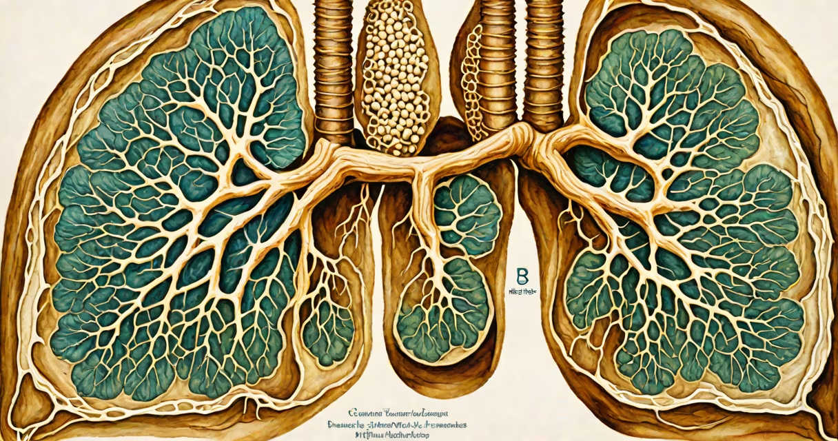
Bronchi and bronchioles are the primary conducting airways in the lungs, responsible for transporting inhaled air from the trachea to the alveoli where gas exchange occurs. They form a branching network that divides progressively into smaller tubes until reaching terminal bronchioles, which lead to respiratory units.
The trachea splits into two main bronchi-one for each lung-at the level of the fifth thoracic vertebra. These main bronchi enter the hilum of each lung and continue dividing into lobar bronchi (three in the right lung, two in the left), then segmental bronchi, and finally into smaller bronchioles. This division follows a pattern of approximately 23 generations, ending at the terminal bronchioles.
Each generation reduces the diameter of the airway. The main bronchi are about 2 cm long and 1.5 cm wide. By the time air reaches the respiratory bronchioles, the diameter is less than 1 mm. This progressive narrowing increases resistance to airflow, but the large surface area of the branching network ensures efficient distribution of air throughout the lungs.
Bronchi and bronchioles maintain airway patency through cartilage rings in the larger airways and smooth muscle in the smaller ones. The cartilage provides structural support, preventing collapse during inhalation. As the airways narrow, cartilage disappears entirely beyond the 10th generation, leaving only smooth muscle and connective tissue.
Smooth muscle in the bronchioles allows for changing regulation of airflow. Contraction narrows the airway (bronchoconstriction), reducing airflow. Relaxation widens it (bronchodilation), increasing airflow. This mechanism is critical during exercise or in response to allergens.
Epithelial lining changes along the pathway. The trachea and bronchi have pseudostratified ciliated columnar epithelium with mucus-producing goblet cells. Mucus traps particles and pathogens, and cilia move them upward toward the pharynx-a process called mucociliary clearance. In bronchioles, the epithelium transitions to simple ciliated columnar and then cuboidal epithelium, with fewer goblet cells and no submucosal glands.
The airway system is organized into distinct zones based on function and structure:
| Level | Diameter (approx.) | Structure | Function |
|---|---|---|---|
| Main bronchus | 15-20 mm | Cartilage rings, ciliated epithelium | Conducts air to lung lobes |
| Lobar bronchus | 6-10 mm | Continued cartilage support | Divides into segments |
| Segmental bronchus | 3-5 mm | Reduced cartilage | Supplies specific lung segments |
| Terminal bronchiole | 0.5-1 mm | No cartilage, smooth muscle only | End of conducting zone |
| Respiratory bronchiole | <0.5 mm | Alveoli begin to appear on walls | Transition to gas exchange |
A key distinction often missed: bronchi are part of the conducting zone, while bronchioles mark the beginning of the respiratory zone. The conducting zone includes all airways from the nose to the terminal bronchioles-its sole purpose is to transport air. The respiratory zone, starting at the respiratory bronchioles, includes structures involved in gas exchange.
Non-obvious fact: Bronchioles lack cartilage and are therefore more prone to collapse under pressure. This makes them vulnerable in conditions like asthma, where inflammation and smooth muscle contraction further reduce diameter. A 50% reduction in bronchiole diameter increases resistance by 16-fold due to Poiseuille's law-airflow drops sharply even with minor narrowing.
Bronchi and bronchioles are essential for delivering oxygen-rich air to alveoli and removing carbon dioxide. Without this network, gas exchange cannot occur efficiently. The entire respiratory system depends on their integrity.
In clinical practice, dysfunction of bronchi and bronchioles leads to significant disease. Chronic obstructive pulmonary disease (COPD) involves chronic inflammation and remodeling of bronchioles, leading to irreversible airflow limitation. Asthma causes reversible bronchoconstriction in small airways, resulting in wheezing and dyspnea. Bronchiectasis is a condition where bronchi become permanently dilated due to infection or inflammation, impairing mucus clearance.
During mechanical ventilation, ventilator-induced lung injury can result from high-pressure breaths causing shear stress on bronchioles. Studies show that excessive tidal volumes (>10 mL/kg) increase the risk of bronchiolar rupture and alveolar damage. The American Thoracic Society recommends limiting tidal volume to 6-8 mL/kg in acute respiratory distress syndrome (ARDS) to protect small airways.
Many assume bronchi and bronchioles serve the same function. In reality, bronchi primarily conduct air, while bronchioles regulate flow and initiate gas exchange. The term "bronchiole" specifically refers to airways smaller than 1 mm in diameter-larger airways are classified as bronchi.
Another misconception: bronchioles do not produce surfactant. Surfactant is synthesized by type II alveolar cells in the alveoli, not in airway epithelium. However, bronchiolar epithelium does secrete a thin layer of fluid that keeps the airway moist and aids in particle trapping.
I once observed a cadaver dissection where a student incorrectly labeled a segmental bronchus as a bronchiole. The error stemmed from misjudging size. The bronchus was 4 mm in diameter-well above the threshold for bronchiole classification. This highlights the importance of anatomical precision: mistaking a bronchus for a bronchiole alters understanding of airflow dynamics and pathology.
Consider a 30-year-old person breathing normally. Inhaled air travels through the trachea, enters the right main bronchus (diameter ~18 mm), then the lobar bronchus (diameter ~8 mm), and continues down to a terminal bronchiole (diameter ~0.7 mm). Total resistance in the airway system averages 0.5-1.0 cm H₂O/L/s in healthy adults.
Now consider the same person during an asthma attack. Inflammatory mediators like histamine cause smooth muscle contraction in bronchioles. A 50% reduction in diameter (from 0.7 mm to 0.35 mm) increases resistance by 16 times-according to Poiseuille's law (resistance ∝ 1/r⁴). This results in audible wheezing and reduced expiratory flow.
Peak expiratory flow rate (PEFR) drops from 500 L/min to around 100 L/min in severe asthma. This demonstrates how small structural changes in bronchioles dramatically affect function. Treatment with inhaled beta-agonists relaxes smooth muscle, restoring diameter and reducing resistance.
Bronchi and bronchioles are not interchangeable terms. Bronchi are larger, cartilaginous, and part of the conducting system. Bronchioles are smaller, muscle-dominated, and begin the transition to gas exchange. Their precise roles ensure that oxygen reaches the bloodstream efficiently and carbon dioxide is expelled.
Understanding bronchi and bronchioles is fundamental to diagnosing and managing respiratory diseases. Their structure enables both air transport and fine control of airflow, making them central to human respiration.