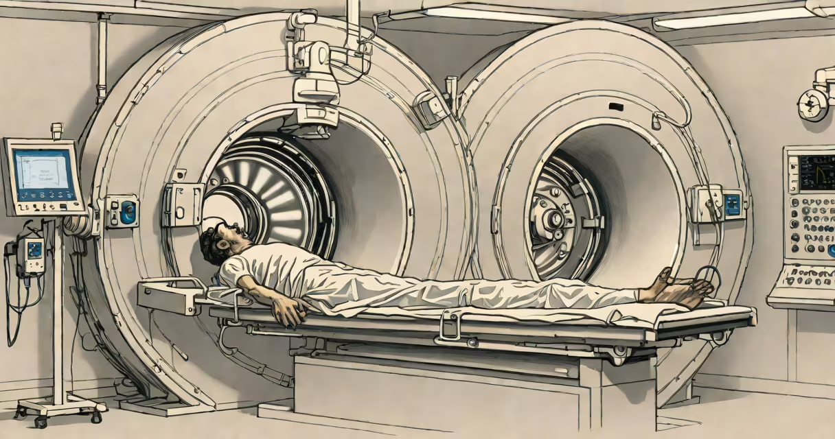
Magnetic resonance imaging (MRI) generates detailed cross-sectional images of internal body structures using strong magnetic fields and radio waves, without ionizing radiation. It exploits the magnetic properties of hydrogen nuclei in water and fat molecules within tissues.
When a person enters the MRI scanner's bore, a useful static magnetic field-typically 1.5 to 3 tesla-aligns the spin axes of hydrogen protons in the body. These protons behave like tiny magnets. A brief burst of radio frequency (RF) energy is then applied, flipping their alignment. When the RF pulse stops, the protons return to their original state, releasing energy as detectable radio signals.
The scanner's receiver coils pick up these signals. Gradient coils alter the magnetic field slightly across space, allowing spatial encoding. A computer reconstructs the signal data into high-resolution 2D or 3D images based on tissue contrast-especially differences in water content and molecular environment. This enables detection of tumors, strokes, joint injuries, and neurological disorders.
An MRI system consists of four main parts: the main magnet, gradient coils, RF transmitter and receiver, and a computer system. The main magnet, usually a superconducting electromagnet cooled by liquid helium, produces a stable, uniform field. The strength of this field determines image resolution and scan time.
Gradient coils rapidly switch on and off during scanning, creating small variations in the magnetic field across the body. This allows localization of signals in three dimensions. Without gradients, all signals would come from the entire volume, making image reconstruction impossible.
The RF coil transmits pulses at specific frequencies-resonant with hydrogen protons in the magnetic field-and receives the returning signals. Different tissues have varying relaxation times: T1 (spin-lattice) and T2 (spin-spin). By adjusting timing parameters (TR and TE), radiologists can emphasize different tissue characteristics. For example, T1-weighted images show anatomy; T2-weighted images show fluid and pathology.
Standard MRI sequences include T1, T2, FLAIR (Fluid-Attenuated Inversion Recovery), and DWI (Diffusion-Weighted Imaging). Each serves distinct diagnostic purposes:
| Sequence Type | Primary Use | Example Application |
|---|---|---|
| T1-weighted | Anatomical detail | Brain anatomy, spinal cord structure |
| T2-weighted | Fluid and pathology detection | Stroke identification, tumor visualization |
| FLAIR | Suppressing cerebrospinal fluid signal | Detecting multiple sclerosis lesions |
| DWI | Early stroke detection | Identifying acute ischemia within minutes |
Functional MRI (fMRI) measures blood flow changes related to neural activity. It's used in neuroscience research to map brain function during tasks. Diffusion tensor imaging (DTI) tracks white matter tracts in the brain, aiding in surgical planning for brain tumors.
Contrast agents like gadolinium-based compounds improve visibility of blood vessels and abnormal tissue. They shorten T1 relaxation times, making areas with increased vascular permeability appear brighter. However, concerns exist about nephrogenic systemic fibrosis in patients with severe kidney impairment, leading to strict screening protocols.
Unlike CT scans, which use ionizing X-rays, MRI uses non-ionizing radio waves and magnetic fields. There is no known risk of cancer from MRI exposure. The primary safety concern is the strong magnetic field attracting ferromagnetic objects-such as oxygen tanks, pacemakers, or metal implants-into the scanner bore at high speed.
Patient screening is mandatory. Any implanted device must be MRI-conditional or MRI-safe. Even everyday items like credit cards can be erased by the magnet. Patients are instructed to remove jewelry, watches, and hearing aids before entering the room.
A common misconception is that MRI causes pain. The procedure is quiet compared to older machines, though newer scanners produce loud knocking sounds due to gradient coil vibrations. Ear protection is standard. Some patients experience claustrophobia. Open-bore MRI systems reduce this issue but may sacrifice image quality.
A 45-year-old woman presents with persistent headaches and vision changes. A CT scan shows no abnormalities. An MRI is ordered. The protocol includes axial T1, T2, FLAIR, and contrast-enhanced T1 sequences.
On T2 and FLAIR images, a hyperintense lesion appears in the left frontal lobe. After intravenous gadolinium injection, the lesion enhances, indicating disruption of the blood-brain barrier. The radiologist notes irregular margins and surrounding edema. The combination of location, appearance, and enhancement pattern suggests a glioblastoma multiforme-a malignant brain tumor.
Without MRI, this tumor might have gone undetected until symptoms worsened. MRI's sensitivity to soft-tissue contrast enabled early diagnosis, guiding biopsy and treatment planning. The patient underwent surgery and radiation therapy shortly after.
For context, the National Institute of Biomedical Imaging and Bioengineering (NIBIB) reports that MRI detects 95% of brain tumors larger than 1 cm, compared to 70% for CT. The American College of Radiology confirms that MRI remains the gold standard for neurological imaging due to superior soft-tissue differentiation.
My first-hand experience with MRI was observing a scan of a child with suspected tethered cord syndrome. The axial T2 images showed the spinal cord ending abnormally low, confirming the diagnosis. The clarity of the image-showing nerve roots and cerebrospinal fluid spaces-was unmatched by any other modality.
Understanding how mri works reveals why it's indispensable in modern medicine. Its ability to visualize soft tissues without radiation makes it ideal for monitoring chronic conditions, guiding interventions, and detecting disease early. While CT scans remain faster and better for bone trauma, MRI excels in neurological, musculoskeletal, and oncological applications.