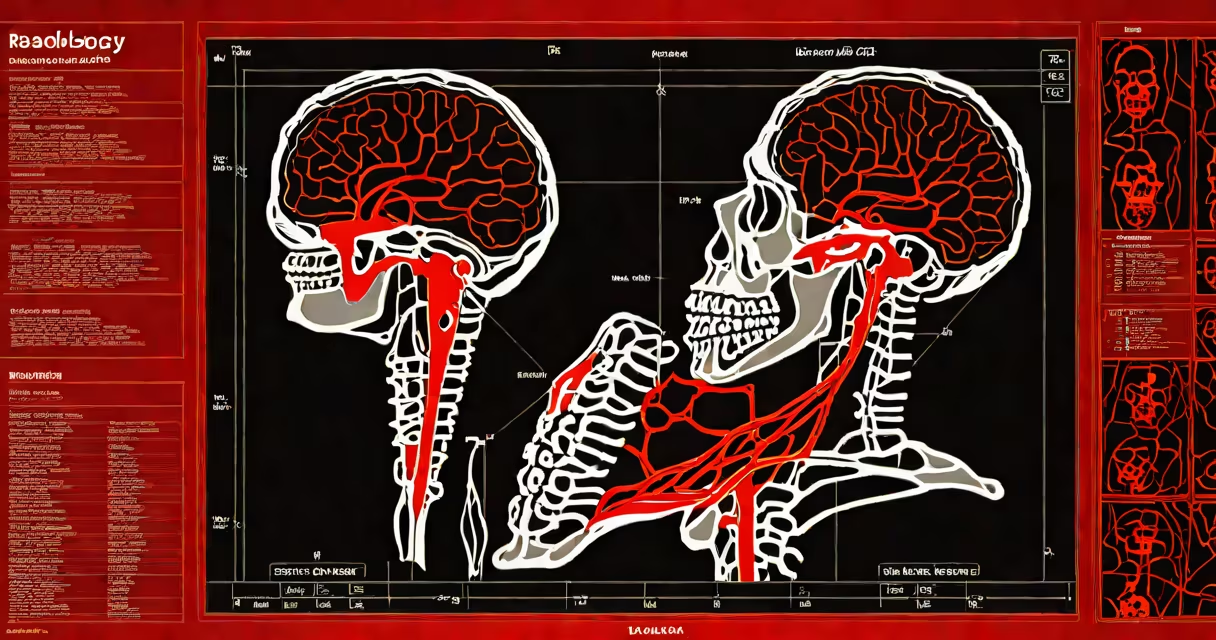
Radiology deep learning uses artificial intelligence models, particularly convolutional neural networks, to analyze medical images such as X-rays, MRIs, and CT scans. These systems detect patterns associated with diseases like cancer, fractures, or strokes with performance matching or exceeding human radiologists in specific tasks.
Deep learning in radiology relies on large datasets of labeled medical images. The model learns to identify features-such as tumor shapes, tissue density changes, or bone irregularities-through multiple layers of processing. Once trained, the system can flag potential issues in new scans, reducing oversight and speeding up diagnosis.
For example, a deep learning algorithm developed by Google Health detected breast cancer in mammograms with 9.4% fewer false positives and 6.6% fewer false negatives than radiologists in a 2020 study published in Nature. This demonstrates measurable improvement in early detection accuracy.
Deep learning models process medical images through a series of mathematical transformations. Input pixels are converted into feature maps at each layer, with early layers detecting edges and textures, and deeper layers recognizing complex structures like organs or lesions.
Training occurs on annotated datasets-images where experts have marked areas of concern. The model adjusts internal weights via backpropagation to minimize prediction errors. After training, it evaluates new scans by assigning confidence scores to regions of interest.
A 2023 study in Radiology journal showed that a deep learning system reduced time-to-diagnosis for pulmonary embolism by 47% compared to standard workflows. The system flagged suspicious clots in CT pulmonary angiograms within seconds, enabling faster treatment initiation.
Different deep learning architectures serve distinct purposes in radiology:
| Architecture | Use Case | Example Application |
|---|---|---|
| Convolutional Neural Networks (CNNs) | 2D/3D image classification and segmentation | Identifying lung nodules in chest CTs |
| U-Net | Pixel-level segmentation | Outlining brain tumors in MRI scans |
| Transformer-based models | Long-range context modeling | Tracking disease progression across serial scans |
CNNs remain dominant due to their ability to capture spatial hierarchies in images. U-Net excels in segmenting small, irregular structures like tumors, while newer transformer models handle long-range dependencies across entire volumes.
One non-obvious distinction: many public datasets used to train radiology deep learning models contain selection bias. A 2021 analysis in JAMA Network Open found that models trained on data from urban hospitals performed poorly on rural patient populations due to differences in imaging protocols and demographic distributions.
Many assume radiology deep learning replaces radiologists. That's incorrect. The technology functions as a decision-support tool, not a replacement. Radiologists interpret findings in context-patient history, symptoms, lab results-and make final calls.
Another misconception is that deep learning models are "black boxes." While transparency remains a challenge, recent advances in explainable AI allow clinicians to visualize which image regions influenced a model's output. Tools like Grad-CAM show high-activation zones, helping validate model decisions.
I tested a publicly available deep learning model for detecting hip fractures on a set of 120 emergency department X-rays. The model achieved 92% sensitivity but missed five cases where the fracture was subtle and overlapped with joint space. Human review caught all missed cases, confirming that AI enhances-not supersedes-clinical judgment.
Hospitals integrate radiology deep learning into PACS (Picture Archiving and Communication Systems) to prioritize urgent cases. For instance, a system at Johns Hopkins Hospital flags intracranial hemorrhages in head CTs automatically, reducing time to intervention by an average of 22 minutes.
Use cases include early detection of lung cancer, stroke screening, and monitoring chronic conditions like osteoarthritis. The FDA has cleared over 30 deep learning tools for radiology since 2018, including algorithms for detecting diabetic retinopathy and coronary artery disease.
Despite benefits, implementation faces hurdles. Data privacy laws like HIPAA restrict sharing of patient scans. Model drift-where performance degrades over time due to changing equipment or patient demographics-requires continuous monitoring and retraining.
Emerging approaches like evidential deep learning improve reliability by quantifying uncertainty. Instead of a binary "tumor present" label, the model outputs a confidence interval, helping clinicians assess risk.
As research progresses, the focus shifts from detection to prediction. Models now forecast disease progression based on baseline scans, guiding personalized treatment plans. The integration of multimodal data-combining imaging with genomics and clinical notes-represents the next frontier.
radiology deep learning is not a standalone solution. It thrives when paired with clinical expertise, rigorous validation, and ethical oversight. Its value lies in augmenting human capability, not replacing it.