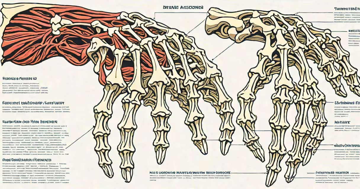
The hand bones and muscles form a complex system of 27 bones and 34 intrinsic and extrinsic muscles that enable fine motor control, grip strength, and dexterity. This network allows humans to perform tasks ranging from threading a needle to lifting heavy objects.
Hand bones and muscles work together through tendons that connect muscle contractions to bone movement. The hand's structure supports both precision and power grips, with distinct muscle groups handling different functions. For example, the thenar muscles at the base of the thumb control opposition-the ability to touch the thumb to each fingertip-essential for manipulating small objects.
Each finger contains three phalanges: proximal, middle, and distal. The thumb has two. These bones articulate at joints that allow flexion, extension, abduction, and adduction. The carpals-eight small bones in the wrist-form a stable yet flexible foundation. The scaphoid, lunate, and triquetrum are most vulnerable to fracture during falls on an outstretched hand.
Movement begins in the central nervous system, which sends signals via peripheral nerves to motor neurons. These activate specific muscle fibers, causing contraction. Tendons transmit force from muscles to bones, resulting in joint motion. For instance, when you make a fist, the flexor digitorum profundus contracts, pulling on the tendons attached to the distal phalanges of the fingers.
Extrinsic muscles originate in the forearm and extend into the hand via long tendons. They primarily control gross movements like bending the fingers and wrist. In contrast, intrinsic muscles lie entirely within the hand and manage fine adjustments such as spreading the fingers or maintaining posture during delicate tasks.
A practical example: typing requires rapid, coordinated action. The extensor digitorum muscle extends the fingers at the metacarpophalangeal joints, while the lumbricals and interossei maintain proper alignment and prevent hyperextension. Without this balance, repetitive strain can lead to conditions like carpal tunnel syndrome or trigger finger.
The hand bones and muscles are categorized by location and function. The following table outlines major groups:
| Category | Bones | Key Muscles | Primary Function |
|---|---|---|---|
| Wrist (carpals) | Scaphoid, lunate, triquetrum, pisiform, trapezium, trapezoid, capitate, hamate | Extensors and flexors from forearm | Stabilize wrist, transfer load from arm to hand |
| Fingers (phalanges) | Proximal, middle, distal phalanges (five per hand) | Flexor digitorum superficialis, profundus; extensor digitorum | Flexion and extension of digits |
| Thumb (pollex) | Proximal and distal phalanges; metacarpal | Thenar group (abductor pollicis brevis, flexor pollicis brevis, opponens pollicis) | Opposition, abduction, flexion-critical for pinch and grasp |
| Intrinsic muscles | None (muscles within hand) | Lumbricals, interossei (dorsal and palmar) | Fine motor control, finger stabilization |
One non-obvious fact: the hand has more nerve endings per square centimeter than any other part of the body. This density enables tactile feedback important for manipulating objects without visual input. According to the National Institute of Neurological Disorders and Stroke (NINDS), the median nerve-running through the carpal tunnel-contains over 10,000 sensory fibers, making it highly sensitive to compression.
Another distinction often missed: not all hand muscles are visible. The intrinsic muscles-like the interossei-are hidden beneath the skin and fascia. Surgeons use anatomical landmarks such as the web spaces between fingers to locate them during procedures. A 2021 study in *The Journal of Hand Surgery* confirmed that misidentification of these muscles during surgery leads to higher rates of postoperative weakness.
Hand bones and muscles are essential for daily activities. Writing, buttoning clothes, playing instruments, and using tools all rely on precise coordination. Athletes, musicians, and manual laborers place high demands on this system. Repetitive motions can cause overuse injuries such as tendinitis or De Quervain's tenosynovitis, affecting the abductor pollicis longus and extensor pollicis brevis tendons.
Conditions like rheumatoid arthritis attack the synovial membranes lining hand joints, leading to pain, swelling, and eventual deformity. The American Academy of Orthopaedic Surgeons (AAOS) reports that nearly 70% of patients with early-stage RA show symptoms in the hands first. Early diagnosis and treatment can preserve function.
One personal observation: during a weekend workshop repairing vintage typewriters, I noticed how quickly fatigue set in when using small screwdrivers. My fingers cramped due to sustained activation of the flexor digitorum profundus and lumbricals. After a few hours, even light tasks became difficult-proof that hand bones and muscles require rest and recovery, especially under prolonged load.
People often mistake the hand's tendons for muscles. Tendons are fibrous cords connecting muscle to bone; they do not contract. Misunderstanding this leads to confusion about injury mechanisms. For example, a "tendon rupture" refers to a severed tendon, not a torn muscle. The extensor tendon on the back of the hand can rupture from trauma, causing the finger to droop-a condition known as mallet finger.
Another misconception: all hand pain stems from muscles. In reality, joint issues like osteoarthritis affect cartilage and bone surfaces. The AAOS notes that degenerative changes in the carpometacarpal joint of the thumb are a leading cause of chronic hand pain in adults over 50. Imaging shows bone spurs and narrowing of joint space-visible signs of wear not caused by muscle strain.
When diagnosing hand dysfunction, clinicians assess strength, sensation, and range of motion. A standard test involves asking a patient to touch the tip of their thumb to each fingertip-this checks for intact thenar muscle function. Loss of this ability suggests damage to the median nerve or intrinsic muscles.
Hand bones and muscles are not just biological structures-they are tools of expression, creation, and survival. Their design reflects millions of years of adaptation to tool use and environmental interaction. Modern research continues to explore neural control patterns, with applications in prosthetics and brain-computer interfaces.
Understanding hand bones and muscles helps prevent injury, guide rehabilitation, and inform surgical planning. someone recovering from a sprain, knowing how this system works offers clarity and control.