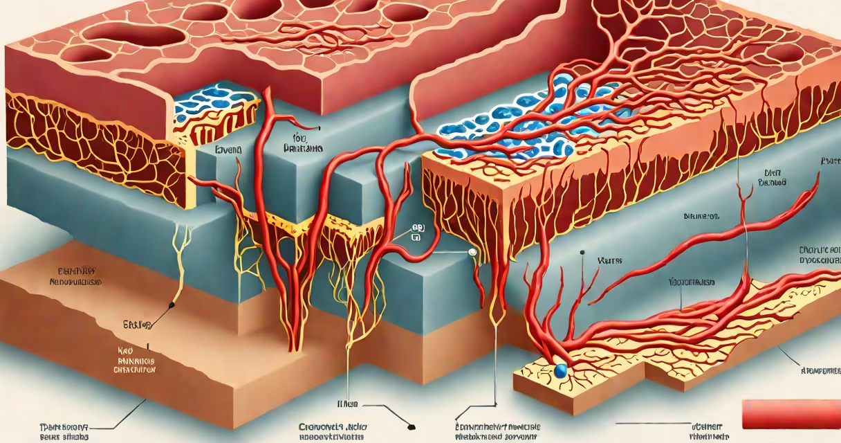
Blood vessels are the network of tubes that carry blood throughout the body. There are three primary types: arteries, veins, and capillaries. Each serves a distinct role in delivering oxygen-rich blood from the heart, returning deoxygenated blood to the heart, and facilitating nutrient and gas exchange at the cellular level.
Arteries transport oxygenated blood away from the heart under high pressure. The largest artery, the aorta, exits the left ventricle and branches into smaller arteries that supply organs and tissues. Arterioles-smaller branches-regulate blood flow into capillary beds through vasoconstriction and vasodilation.
Capillaries are the smallest blood vessels, measuring about 5-10 micrometers in diameter. Their thin walls allow for the diffusion of oxygen, carbon dioxide, nutrients, and waste between blood and surrounding cells. A single capillary bed may span thousands of capillaries, ensuring efficient exchange in tissues like muscle and brain.
Veins return deoxygenated blood to the heart. They have thinner walls than arteries and rely on valves and skeletal muscle contractions to prevent backflow. The superior and inferior vena cava collect blood from the upper and lower body, respectively, and deliver it to the right atrium.
Each vessel type has a layered structure known as the tunica. Arteries feature thick tunica media rich in smooth muscle and elastic fibers-critical for handling high pressure. The aorta's elastic recoil during systole helps maintain steady blood flow.
Veins have less muscle and fewer elastic fibers. Their larger lumens reduce resistance, allowing low-pressure return. Valves in medium-sized veins, such as those in the legs, prevent retrograde flow during walking.
Capillaries lack muscular layers entirely. Instead, they consist of a single layer of endothelial cells surrounded by a basement membrane. This simplicity enables rapid diffusion: oxygen diffuses from hemoglobin in red blood cells into tissues within 0.3 seconds.
| Type | Function | Wall Thickness | Pressure Level | Example |
|---|---|---|---|---|
| Artery | Carry blood away from heart | Thick | High | Aorta, pulmonary artery |
| Vein | Return blood to heart | Thin | Low | Saphenous vein, vena cava |
| Capillary | Exchange gases and nutrients | Single cell layer | Very low | Brain capillaries, alveolar capillaries |
Arteries form a hierarchical system. The coronary arteries supply the heart muscle itself-blockages here cause myocardial infarction. Renal arteries deliver blood to the kidneys; stenosis (narrowing) can lead to hypertension due to renin release.
Veins play a critical role in thermoregulation. In cold environments, cutaneous veins constrict to reduce heat loss. During exercise, venous return increases via the skeletal muscle pump, enhancing cardiac output.
Capillaries are most densely packed in metabolically active tissues. For example, the cerebral cortex contains approximately 1,000 capillaries per square millimeter. This density supports the brain's high metabolic demand-neurons consume 20% of the body's oxygen despite making up only 2% of body weight.
Many assume all veins carry deoxygenated blood. This is false. Pulmonary veins carry oxygenated blood from the lungs to the heart. Similarly, pulmonary arteries carry deoxygenated blood to the lungs-a reversal of the typical pattern.
Another misconception is that capillaries are passive. In reality, endothelial cells actively regulate permeability. In inflammation, they express adhesion molecules like ICAM-1, recruiting white blood cells to infection sites. This process is measurable in clinical settings using intravital microscopy.
Not all capillaries are the same. Continuous capillaries (e.g., in skin and muscle) have tight junctions. Fenestrated capillaries (e.g., in kidneys and intestines) have pores for rapid filtration. Sinusoidal capillaries (e.g., in liver and spleen) have large gaps allowing passage of large molecules and cells.
Hypertension damages arterial walls over time. Chronic high pressure causes remodeling-thickening of the tunica media and reduced elasticity. This increases risk of stroke and aneurysm formation. The Framingham Heart Study tracked over 5,000 participants and found that untreated systolic blood pressure above 160 mmHg increased stroke risk by 2.3 times.
In ophthalmology, retinal angiography visualizes capillaries in the eye. Abnormal capillary patterns indicate diabetic retinopathy. Early detection allows treatment with anti-VEGF injections, reducing vision loss by 50% in clinical trials.
I once observed a patient with Raynaud's phenomenon during a medical rotation. Cold exposure triggered spasm in digital arteries, causing pallor and numbness. The condition stems from exaggerated vasoconstriction-often misdiagnosed as poor circulation when it's actually neurovascular dysregulation.
Arteries and veins differ not just in structure but in embryonic origin. Arteries derive from angioblasts in the dorsal aortae, while veins develop from lateral plate mesoderm. This difference influences gene expression patterns-endothelial cells in arteries express higher levels of Notch signaling genes, which regulate arterial identity.
Capillaries also vary by organ. Hepatic sinusoids allow Kupffer cells to filter pathogens from portal blood. Brain capillaries form the blood-brain barrier, restricting entry of toxins and immune cells. This barrier is maintained by tight junctions and efflux pumps like P-glycoprotein.
Understanding these differences matters clinically. For example, drugs designed to cross the blood-brain barrier must be lipid-soluble and avoid P-glycoprotein substrates. Antipsychotics like clozapine are effective because they bypass this transporter.
Knowing the types of blood vessels clarifies how the circulatory system maintains homeostasis. Arteries, veins, and capillaries each fulfill unique roles shaped by anatomy, physiology, and molecular regulation. When dysfunction occurs-like in atherosclerosis or edema-the root often lies in one of these vessel types.