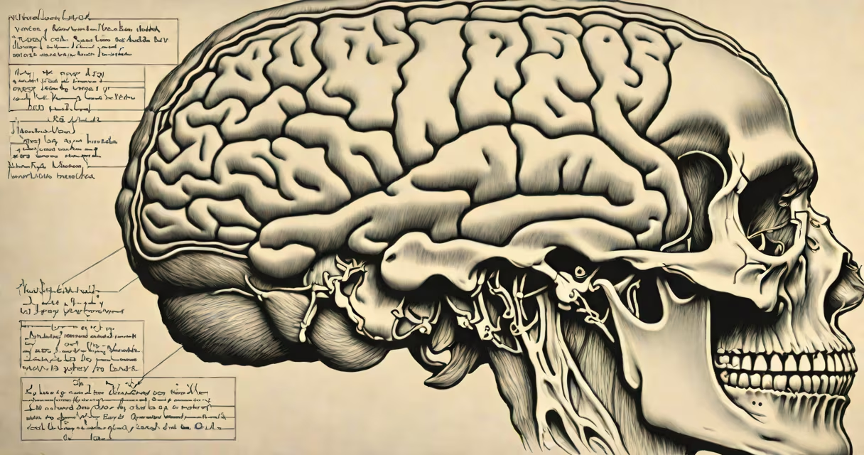
A glioma tumor is a primary brain or spinal cord cancer that arises from glial cells-supportive cells in the central nervous system. These tumors are classified by cell type, location, and malignancy grade, with grades ranging from I (least aggressive) to IV (most aggressive). Gliomas account for approximately 30% of all primary brain tumors and 80% of malignant ones, according to the American Brain Tumor Association.
Gliomas originate in astrocytes, oligodendrocytes, or ependymal cells. The most common subtype, glioblastoma (GBM), develops from astrocytes and is classified as a grade IV glioma under the World Health Organization's (WHO) 2021 classification system. This system integrates histological features with molecular markers such as IDH mutation status and 1p/19q co-deletion, which influence prognosis and treatment.
Unlike metastatic tumors that spread from other organs, gliomas begin within the brain or spinal cord. They grow slowly in low-grade forms but infiltrate surrounding tissue rapidly in high-grade variants. Symptoms depend on location-frontal lobe gliomas may cause personality changes, while temporal lobe tumors often lead to seizures. Early detection improves outcomes, though median survival for grade IV gliomas remains around 12 to 15 months despite treatment.
Gliomas form when glial precursor cells undergo genetic mutations that disrupt normal growth control. Common alterations include mutations in TP53, PTEN, and EGFR genes, especially in glioblastoma. These changes allow uncontrolled proliferation and resistance to apoptosis, enabling tumor progression.
The WHO classification system now combines histopathology with molecular diagnostics. For example, a tumor diagnosed as "astrocytoma, IDH-mutant, grade 4" indicates a high-grade tumor driven by an IDH gene mutation. This contrasts with "astrocytoma, IDH-wildtype, grade 4," which behaves more aggressively and is often referred to as glioblastoma, IDH-wildtype. The distinction affects both prognosis and therapeutic decisions.
Imaging plays a key role in initial assessment. MRI scans typically show contrast enhancement in high-grade gliomas due to blood-brain barrier breakdown. A typical finding is a heterogeneous mass with necrosis and surrounding edema-known as the "ring-enhancing lesion." However, imaging alone cannot confirm diagnosis; biopsy remains essential for definitive classification.
Gliomas are categorized into four main types based on cell origin and behavior:
| Type | Cell Origin | Common Grade | Prognosis | Key Features |
|---|---|---|---|---|
| Diffuse Astrocytoma | Astrocytes | I-III | Median survival: 5-10 years (grade III) | IDH mutation common; slow growth |
| Oligodendroglioma | Oligodendrocytes | II-III | Median survival: 10-15 years (grade III) | 1p/19q co-deletion present; responsive to chemotherapy |
| Ependymoma | Ependymal cells | I-III | Variable; often better outcome than astrocytomas | Often located in posterior fossa; may recur |
| Glioblastoma (GBM) | Astrocytes | IV | Median survival: 12-15 months | Highly invasive; necrosis and microvascular proliferation |
Grade I gliomas are rare and usually curable with surgery alone. Grade II tumors are slow-growing but tend to progress over time. Grades III and IV represent increasingly aggressive disease. Grade IV gliomas-especially GBM-are among the most challenging cancers to treat due to their diffuse infiltration and resistance to therapy.
One non-obvious fact: not all gliomas require immediate treatment. Low-grade gliomas (grade II) may be monitored via serial MRI scans if asymptomatic. The European Organisation for Research and Treatment of Cancer (EORTC) trial 22844 showed that observation delayed intervention without reducing survival in selected patients.
Many people assume all brain tumors are equally dangerous. In reality, gliomas vary widely in behavior. A grade I pilocytic astrocytoma in a child's cerebellum may be cured with complete surgical removal and have a 95% long-term survival rate. In contrast, a grade IV glioblastoma in an adult has a five-year survival rate below 10%, despite aggressive multimodal therapy.
Another misconception is that gliomas are always fatal. While high-grade gliomas remain difficult to cure, advances in immunotherapy and targeted treatments offer hope. For example, the drug temozolomide, used in combination with radiation, extends survival in GBM patients by about 2.5 months compared to radiation alone, per a 2016 study published in *The New England Journal of Medicine*.
Some patients confuse gliomas with benign brain masses like meningiomas. Meningiomas arise from meningeal cells, not glial cells, and are typically slow-growing with lower recurrence rates. Distinguishing between them requires imaging and pathology-misdiagnosis can delay proper care.
A 52-year-old woman presented with worsening headaches and left-sided weakness. An MRI revealed a 4.5 cm mass in the right frontal lobe with ring enhancement and surrounding edema. After biopsy, pathology confirmed a glioblastoma, IDH-wildtype, with hyperexpression of EGFR and loss of PTEN. Molecular testing showed no 1p/19q co-deletion.
She underwent maximal safe resection followed by concurrent temozolomide and radiation (Stupp protocol). Post-treatment, her symptoms improved. Repeat MRI at six weeks showed a 60% reduction in tumor volume. She then received six cycles of adjuvant temozolomide. Despite this, progression occurred at 14 months. At that point, she was enrolled in a clinical trial evaluating a personalized neoantigen vaccine.
This case illustrates how diagnosis hinges on both imaging and molecular profiling. The absence of 1p/19q co-deletion ruled out oligodendroglioma, and the IDH-wildtype status indicated a poor prognosis. Yet treatment options extended beyond standard care, including experimental therapies.
When managing a glioma tumor, clinicians consider patient performance status, age, and comorbidities. The Karnofsky Performance Status scale is routinely used to assess functional ability. Patients with scores above 70 are more likely to tolerate aggressive treatment.
Early diagnosis and multidisciplinary evaluation are important. Neuro-oncologists, radiologists, pathologists, and rehabilitation specialists collaborate to tailor care. Genetic counseling may be recommended for families with inherited syndromes linked to glioma risk, such as Li-Fraumeni syndrome or Turcot syndrome.
Understanding what is a glioma tumor involves recognizing its biological complexity, diagnostic precision, and evolving treatment field. With ongoing research, survival rates continue to improve, particularly for subtypes with favorable molecular profiles.