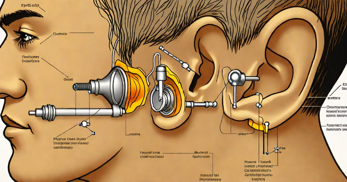
A healthy eardrum, or tympanic membrane, appears translucent, slightly concave, and ranges from pale pink to grayish-white. It features visible landmarks such as the umbo (center point), handle of the malleus (ear bone), and light reflex. Any significant redness, bulging, or discoloration may indicate infection or injury.
The eardrum separates the outer ear from the middle ear. It vibrates in response to sound waves, transmitting those vibrations to tiny bones inside the middle ear. Its structure is delicate-made of three layers: outer skin, fibrous connective tissue, and inner mucous membrane. Damage to any layer can affect hearing and increase infection risk.
Doctors examine the eardrum using an otoscope-a handheld device with a light and magnifying lens. During a routine exam, they assess color, shape, transparency, and movement. A normal eardrum moves slightly when air pressure changes, such as during swallowing.
A healthy eardrum is thin, semi-transparent, and gently curved inward toward the middle ear. This inward curve helps maintain proper pressure balance. The surface is smooth without holes, blisters, or discharge. Color varies by individual but typically falls within a pale pink to grayish-white spectrum.
Key anatomical features include:
When examined under an otoscope, a normal eardrum shows no signs of fluid behind it. Fluid buildup often causes the membrane to appear cloudy or bulge outward. This condition, known as otitis media, affects about 75% of children at least once before age five, according to the American Academy of Pediatrics.
Changes in eardrum appearance signal underlying issues. Redness or intense pink coloring suggests acute infection or inflammation. A swollen, bulging eardrum indicates fluid or pus accumulation-common in middle ear infections. If the eardrum appears yellow or green, it may signal chronic infection or a perforation.
Perforated eardrums show visible holes or tears. They can result from trauma, sudden pressure changes (like during scuba diving), or untreated infections. A perforation reduces hearing ability and increases the risk of further infection. Research from the National Institute on Deafness and Other Communication Disorders notes that most small perforations heal within weeks without surgery.
Chronic conditions like otosclerosis-abnormal bone growth around the eardrum-can cause stiffness and reduced mobility. Over time, this leads to progressive hearing loss. In rare cases, persistent ear pain with a grayish or blackish film may indicate fungal infection, especially in people with weakened immune systems.
Many assume a healthy eardrum should be completely white or shiny. That's incorrect. A normal eardrum is not reflective like glass. It's translucent, allowing faint visibility of structures behind it, such as the ossicles (middle ear bones).
Another myth is that wax buildup distorts the eardrum's appearance. While earwax (cerumen) accumulates in the ear canal, it doesn't touch the eardrum unless pushed too far. Excessive cleaning with cotton swabs can damage the eardrum or push wax deeper, increasing risk of impaction.
Some patients worry about "holes" seen in images online. These are often mislabeled photos or illustrations of diseased ears. A true hole in the eardrum is a medical concern requiring evaluation. If you suspect a perforation, avoid water exposure and see a specialist.
See a healthcare provider if you notice:
Children are especially prone to middle ear infections. Signs include irritability, pulling at the ear, difficulty sleeping, and fever. The American Academy of Pediatrics recommends prompt evaluation when symptoms persist beyond 48 hours.
I once observed a patient with a perforated eardrum during a clinic visit. The hole was about 2 mm wide, located near the upper edge. The patient reported a history of loud noise exposure and recent ear-popping during a flight. After imaging and referral to an ENT specialist, the perforation healed within six weeks with conservative management.
| Feature | Healthy Eardrum | Abnormal Eardrum |
|---|---|---|
| Color | Pale pink to grayish-white | Red, yellow, green, or blackish |
| Transparency | Translucent, allows viewing of internal structures | Cloudy, opaque, or covered in discharge |
| Shape | Slightly concave, smooth | Bulging, sunken, or torn |
| Light Reflex | Visible triangle in front lower quadrant | Missing or distorted |
| Fluid Behind Membrane | None | Present (indicates infection or allergy) |
For context, a normal eardrum is roughly 1 cm in diameter and 0.1 mm thick-about the size of a dime and thinner than a sheet of paper. Its resilience comes from layered construction: outer epithelium, middle fibrous layer, and inner mucosal lining.
Understanding normal eardrum appearance supports early detection of ear problems. Regular check-ups and awareness of changes in hearing or ear sensation improve outcomes. If you have concerns, don't wait-seek evaluation.