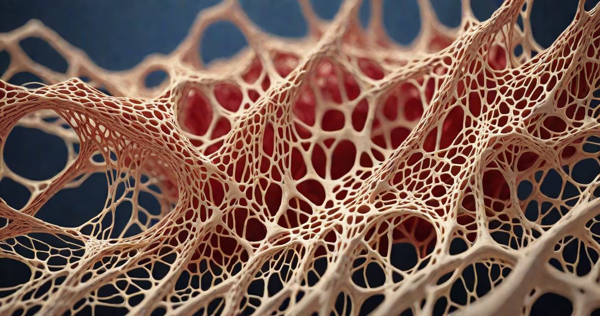
Connective tissue is composed primarily of collagen fibers, elastin proteins, and specialized cells like fibroblasts, embedded in a gel-like extracellular matrix. This structure provides strength, elasticity, and support across organs, bones, and skin.
Connective tissue forms the body's internal scaffolding. It binds structures together, cushions organs, and enables movement. Unlike epithelial or muscle tissues, connective tissue is not tightly packed. Its loose, porous architecture allows nutrients and waste to diffuse easily through the matrix.
The extracellular matrix-about 70% of connective tissue volume-is synthesized by fibroblasts. These cells secrete collagen and elastin, which form long, cross-linked fibers. Collagen gives tensile strength; elastin allows stretch and recoil. Without these proteins, tissues would fail under mechanical stress.
Three elements define connective tissue: structural proteins, cellular elements, and the ground substance. Together, they create a resilient yet flexible structure.
Collagen: The most abundant protein in the human body. Type I collagen makes up 90% of connective tissue. It forms fibrils that resist stretching. A single collagen molecule is 300 nanometers long-long enough to span several cells. In tendons, collagen fibers align in parallel bundles, allowing force transmission during movement.
Elastin: Found in tissues needing rebound, such as lung alveoli and arterial walls. Elastin forms a network of cross-linked chains that return to shape after deformation. A single elastin fiber can stretch to twice its resting length and snap back without damage.
Fibroblasts: The primary cell type in connective tissue. They reside in the matrix and produce both collagen and elastin. When injury occurs, fibroblasts migrate to the site and begin rebuilding the matrix. In wound healing, they differentiate into myofibroblasts-cells that contract to close wounds.
| Component | Function | Example Location |
|---|---|---|
| Collagen (Type I) | Provides tensile strength | Tendons, skin, bone |
| Elastin | Enables elastic recoil | Lungs, arteries, ear cartilage |
| Fibroblasts | Synthesize matrix proteins | Wound sites, dermis, tendons |
Connective tissue is classified by density, fiber type, and function. The most common types include loose, dense regular, dense irregular, and specialized forms like cartilage and bone.
Loose connective tissue-found beneath the skin and around blood vessels-contains few fibers and many fibroblasts. It acts as a cushion and nutrient exchange zone. Adipose tissue, a subtype, stores energy in lipid-filled adipocytes.
Dense regular connective tissue arranges collagen fibers in parallel lines. Tendons and ligaments use this structure to transmit force efficiently. A single tendon can withstand forces exceeding 500 newtons-equivalent to lifting a 50 kg weight.
Dense irregular connective tissue has randomly oriented fibers. It resists multidirectional stress. The dermis layer of skin contains this type, protecting against shear and impact.
Cartilage and bone are specialized connective tissues. Cartilage contains chondrocytes suspended in a firm matrix rich in collagen and proteoglycans. Bone adds mineral salts-calcium phosphate-to harden the matrix. A femur can support over 1,000 kilograms of compressive load due to this mineralization.
Many assume connective tissue is passive or inert. In reality, it is metabolically active. Fibroblasts constantly remodel the matrix in response to mechanical load and injury signals.
Another error is equating all connective tissue with scar tissue. While scars involve fibroblast activity and collagen deposition, normal connective tissue maintains homeostasis. Scar tissue forms after trauma and lacks the organized fiber alignment seen in healthy tissue.
A less obvious point: the extracellular matrix is not just a scaffold. It regulates cell behavior. Matrix stiffness influences whether fibroblasts remain quiescent or activate into repair mode. Soft matrices promote stem cell differentiation into fat cells; stiff ones favor bone formation.
Consider a rotator cuff tear in the shoulder. The supraspinatus tendon-a dense regular connective tissue-fails under repetitive strain. MRI shows a disruption in the aligned collagen fibers. Surgical repair involves reattaching the tendon to the humerus using suture anchors.
During recovery, fibroblasts infiltrate the area and deposit new collagen. Over six months, the matrix reorganizes. However, the repaired tendon never regains full mechanical strength. Studies show it retains only 60-70% of original tensile strength, highlighting the limits of natural repair.
This example shows connective tissue's role in biomechanics and healing. It also reveals why injuries to tendons and ligaments take longer to heal than muscle strains-they depend on slow matrix remodeling.
I once observed a patient with Ehlers-Danlos syndrome, a genetic disorder affecting collagen synthesis. Their skin stretched excessively and joints dislocated easily. Blood tests confirmed low levels of type III collagen. This condition illustrates how a single protein deficiency disrupts entire tissue systems.
Abnormalities in connective tissue components lead to disease. Marfan syndrome results from mutations in the FBN1 gene, which codes for fibrillin-a protein that regulates elastin assembly. Patients develop elongated limbs, heart valve defects, and vision problems due to weakened connective tissues.
Understanding what connective tissue is made of explains how the body holds itself together. From supporting the spine to enabling joint motion, its composition determines resilience, repair capacity, and susceptibility to disease.